Pancreatic Cancer Ct Scan / Anatomic Definitions of Borderline Resectable Pancreatic ... : For example, the relationship of the cancer in the pancreas to important blood vessels adjacent to the pancreas can be seen, a feature used by surgeons to determine if the.
byAdmin-
0
Pancreatic Cancer Ct Scan / Anatomic Definitions of Borderline Resectable Pancreatic ... : For example, the relationship of the cancer in the pancreas to important blood vessels adjacent to the pancreas can be seen, a feature used by surgeons to determine if the.. Why aren't pancreatic ct scans used in routine physical examinations to detect and prevent the spread of pancreatic cancer? Ct scans can also be used to guide a biopsy needle into a suspected pancreatic tumor. Ct scans can show pancreatitis or pancreatic cancer. Ct scans are often performed to monitor patients after treatment to determine whether the cancer has recurred, changed in size or metastasized this test is not as specific as ct scanning and is not used alone to diagnose pancreatic cancer. First line investigations ct scan for the scanner is shaped like a doughnut.
A contrast dye injected at the start of the here are some other challenges to finding pancreatic cancer early: It takes pictures from different angles. Many cancer centers use a special ct scan method called a pancreatic protocol ct scan. Pancreatic cancer — overview covers symptoms, risk factors, prevention, diagnosis, surgery, chemotherapy and other treatment for cancer of several types of growths can occur in the pancreas, including cancerous and noncancerous tumors. Ct scans create pictures of your pancreas, gallbladder, and bile ducts.
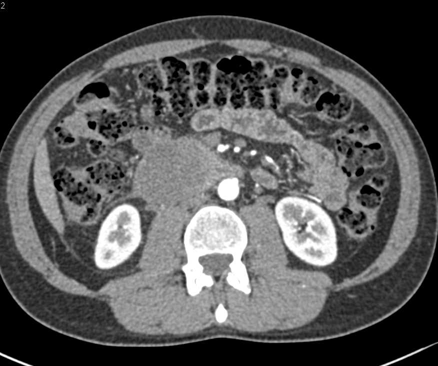
Pancreatic Cancer with Tumor Spread - Pancreas Case ... from ctisus.com Mar 07, 2019 [электронный 24. The most common type of cancer that forms in the. A precontrast scan of the pancreas can be performed to look for calcifications within the pancreas, which may indicate the presence of a focal pancreatitis. The ct scans, four of them over the course of the 11 weeks from my initial appointment with my primary (23 march). Pancreatic cancer is usually staged following a ct scan.29 the most widely used cancer staging system for pancreatic cancer is the one formulated by the american joint committee on cancer (ajcc) together with the union for international cancer control (uicc). The remaining number can be divided into exocrine tumours (such as pancreatic cystic carcinoma) and endocrine tumours (derived from islet cells of the pancreas). From my experience as the patient being diagnosed…. How do ct scans work?
Learn how this test works, as well as its benefits and risks.
Pancreatic cancer will typically refer to ductal carcinoma of the pancreas, which comprises up to 90% of primary pancreatic malignancies. Contrast is a special dye used to make it easier. The abdominal ct scan may show problems with the gallbladder, liver, or pancreas, including Ct scans can also be used to guide a biopsy needle into a suspected pancreatic tumor. Ct stands for computed tomography. Ct scanning will visualize the vast majority of pancreatic cancers, and it provides important information to guide treatment. Ct scans help doctors diagnose and treat medical conditions such as pancreatic cancer. A precontrast scan of the pancreas can be performed to look for calcifications within the pancreas, which may indicate the presence of a focal pancreatitis. Pancreatic cancer (cancer of the pancreas) mainly occurs in people aged over 60. From my experience as the patient being diagnosed…. If you have jaundice and suspected pancreatic cancer, or if you have been diagnosed with pancreatic cancer that is contained within the pancreas (localised pancreatic cancer) after a ct scan, you should also. A ct scan is one of the most common tests for pancreatic cancer. Pet/ct fusion scan enhances ct staging in patients with pancreatic.
A ct scan is one of the most common tests for pancreatic cancer. You will be asked to lie still on a table, which slides into the. A ct scan is often used as the primary test to diagnose or confirm pancreatic cancer. For evaluating possible pancreatic cancer, a multiphase helical ct scan or pancreatic protocol ct scan is often recommended. During an ultrasound, gel is spread over your abdomen and a scanner creates pictures of your organs.
Unresectable Pancreatic Adenocarcinoma: Eight Years Later ... from www.wjon.org An abdominal ct scan is an imaging method. This method focuses on taking pictures of the pancreas at specific. A precontrast scan of the pancreas can be performed to look for calcifications within the pancreas, which may indicate the presence of a focal pancreatitis. The remaining number can be divided into exocrine tumours (such as pancreatic cystic carcinoma) and endocrine tumours (derived from islet cells of the pancreas). Why aren't pancreatic ct scans used in routine physical examinations to detect and prevent the spread of pancreatic cancer? A contrast dye injected at the start of the here are some other challenges to finding pancreatic cancer early: Ct scanning will visualize the vast majority of pancreatic cancers, and it provides important information to guide treatment. Mar 07, 2019 [электронный 24.
The images often help doctors find out if they can remove the tumor through surgery or if it has metastasized (spread to other.
Contrast is a special dye used to make it easier. Computed tomography (ct or cat) scan. The images often help doctors find out if they can remove the tumor through surgery or if it has metastasized (spread to other. Abdominal ct scan can provide a more detailed analysis of the pancreas and other abdominal organs and is much more accurate than ultrasound in detecting pancreatic cancer, with a sensitivity of about 90%. Many cancer centers use a special ct scan method called a pancreatic protocol ct scan. Ct scanning will visualize the vast majority of pancreatic cancers, and it provides important information to guide treatment. Home about pancreatic cancer diagnosis of pancreatic cancer how is pancreatic cancer diagnosed? How do ct scans work? First line investigations ct scan for the scanner is shaped like a doughnut. Learn how this test works, as well as its benefits and risks. Mar 07, 2019 [электронный 24. It can show the pancreas it also helps doctors know how extensively cancer has spread in the pancreas. Before the scan, dye is injected into a vein to help make the pictures clearer.
Pancreatic cancer (cancer of the pancreas) mainly occurs in people aged over 60. Pancreatic cancer will typically refer to ductal carcinoma of the pancreas, which comprises up to 90% of primary pancreatic malignancies. It takes pictures from different angles. A special type of ct known as a multiphase ct scan or pancreatic. An abdominal ct scan is an imaging method.
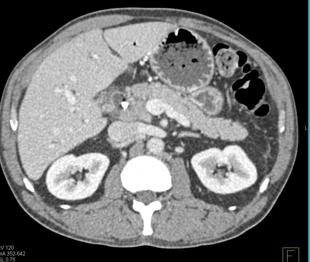
Pancreatic Cancer with Normal Vascular Map - Pancreas Case ... from ctisus.com Hongwei li1, kanru lin3, maximilian reichert2, lina xu1, rickmer braren2 1 introduction. During an ultrasound, gel is spread over your abdomen and a scanner creates pictures of your organs. Computed tomography (ct or cat) scan. A contrast dye injected at the start of the here are some other challenges to finding pancreatic cancer early: Unless other factors make its use unsuitable, a ct scan optimized for imaging the pancreas is the primary option for diagnosis and staging of pancreatic. A ct scan is one of the most common tests for pancreatic cancer. It's difficult to see or feel pancreatic tumors because the pancreas is so. From my experience as the patient being diagnosed….
It's difficult to see or feel pancreatic tumors because the pancreas is so.
Learn how this test works, as well as its benefits and risks. The most common type of cancer that forms in the. Ct stands for computed tomography. Multidetector ct scanning (mdct) using a pancreas protocol is at least as accurate as eus in the overall determination of the resectability of pancreatic algorithm for evaluation of a patient with suspected pancreatic cancer. Ct scans are usually done at a hospital or a radiology clinic. A pet scan is often done in combination with a ct scan. Many cancer centers use a special ct scan method called a pancreatic protocol ct scan. An abdominal ct scan is an imaging method. Before the scan, dye is injected into a vein to help make the pictures clearer. But if a needle biopsy is needed, most doctors prefer to use it might be especially useful for spotting cancer that has spread beyond the pancreas and wouldn't be treatable by surgery. This provides a series of images from many different angles. You will be asked to lie still on a table, which slides into the. It can still miss some small tumours, especially those <3cm.
The remaining number can be divided into exocrine tumours (such as pancreatic cystic carcinoma) and endocrine tumours (derived from islet cells of the pancreas) pancreatic cancer. First line investigations ct scan for the scanner is shaped like a doughnut.